 |
Macular Hole |
 |

Macular Hole |
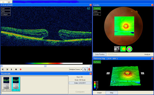
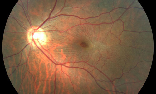
The macula is the portion of the retina responsible for clear, detailed vision. A macular hole is a defect in the central portion of the retina. There are two types of macular hole: Lamellar Macular Hole Full Thickness Macular Hole |
|
Both types of macular holes can occur due to normal vitreous shrinkage that happens as you age, or may occur in those who are nearsighted or have had an injury to the eye. As you age, the vitreous gel shrinks and separates from the macula. Usually, there is little effect on your vision. However, sometimes the gel sticks to the macula and the vitreous is unable to pull away. As the gel pulls on the macula, it begins to stretch. Over time, this stretching of the macula may cause a hole to develop. On rare occasions, a hole may be caused by an injury, or long term swelling of the retina. |
|
Early symptoms of a Macular Hole include blurry or distorted central vision. You might describe it as “fuzzy” or “hazy”. Everyday activities such as reading may become difficult. The words in a book may be distorted. More advanced symptoms include a blind spot in your central vision, and impaired vision both at distance and close range. |

|
These conditions can be diagnosed by a complete eye examination and special photographs called optical coherence photography and a fluorescein angiogram. These tests will determine the depth of the hole and the thickness of the macula around it. |
|
Usually there is no treatment required for a lamellar macular hole; in most cases these close on their own. Full thickness macular holes require Vitrectomy surgery to repair the macular hole and possibly improve your vision. The surgery is done under a microscope. Tiny instruments are used to carefully repair the hole and remove the vitreous completely. The eye is then filled with a special gas bubble to help flatten the macular hole and surrounding retina. The day of surgery, your eye will be patched. You will leave the patch on overnight. You will return to the office the following day to remove the patch and be examined by the doctor. It is normal to have some discomfort after surgery. You will be prescribed eye drops to use after surgery, and given instructions about post-operative care and activities. You must stay in a face-down position after surgery so the gas bubble applies pressure to the macula. Since the retina is the back wall of the eye, you must have your face parallel to the floor to force the gas bubble to press against the retina. The pressure from the gas bubble helps to flatten the retina. The success of your surgery is often dependant on your ability to maintain this position. You will be allowed to sleep on your side, but not on your back while the gas bubble is present in your eye. The gas bubble will slowly dissolve until it disappears completely, which usually takes 10-12 weeks. As the macular hole closes, your vision may slowly start improving. The amount of visual recovery varies for each person. Factors such as the size and depth of the hole, how long it was present prior to surgery and the quality of face down positioning will make your outcome unique to you. The eye takes at least 6 weeks to heal from the surgery itself (90%), and the vision may gradually continue to improve for up to 6 months after surgery (10%). |
|
There is no known way of preventing a macular hole from developing. Therefore, regular eye exams are important to make sure that no new eye conditions are developing. |
|
 |
 |
|
