 |
Branch Retinal Vein Occlusion |
 |

Branch Retinal Vein Occlusion near Fort Myers | ||
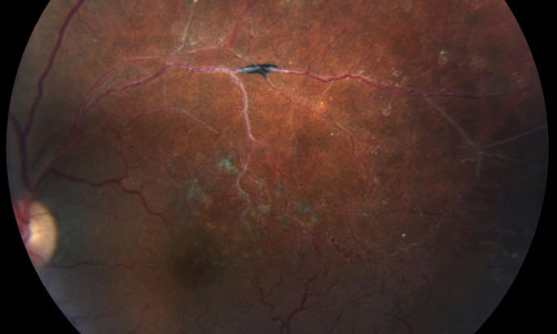
A branch retinal vein occlusion may happen when a thickened artery presses against an underlying vein wall, and a small piece of cholesterol plaque or a small blood clot that has passed through the central retinal vein, becomes lodged in a smaller vein in the eye. It is considered a small stroke in the eye. | ||
| ||
This is most commonly caused by high blood pressure, coronary artery disease, diabetes or vascular disease, however, may also be a complication of other systemic disorders. | ||
| ||
You may notice a sudden loss of peripheral vision in one eye. You may not notice any symptoms in some cases. | ||
| ||
A branch retinal vein occlusion is diagnosed by retinal examination. Special pictures called a fluorescein angiogram, and an OCT (optical coherence tomography) scan will be used to determine the impact on blood flow and the amount of damage caused. You may be asked to have lab tests, a carotid ultrasound and echocardiogram. You will need to also see your medical doctor for a cardiovascular evaluation. | ||
| ||
The treatment for a branch retinal vein occlusion may include injections of medication into the eye, and in rare cases, laser treatment. You will need to have a thorough examination with your medical doctor. The prognosis is usually fairly good, resulting in only a small amount of vision loss. | ||
| ||
It is important to treat the underlying medical causes of your vascular disorder to prevent further retinal vein occlusions. | ||
| ||
 |
RETINA FORT MYERS |
 |
|
