 |
Branch Retinal Artery Occlusion |
 |

Branch Retinal Artery Occlusion near Fort Myers | ||
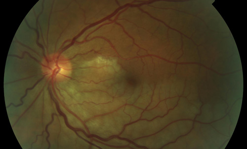
A branch retinal artery occlusion usually occurs at the junction of two smaller arteries, where blood flow is disrupted and an area of ischemia occurs. | ||
| ||
The cause of a branch retinal artery occlusion is often caused by a plaque, calcium, or platelet deposit becoming lodged at an arterial bifurcation in the eye. This is sometimes preceded or followed by a stroke or TIA. Most people have some degree of permanent vision loss as a result of this condition. | ||
| ||
Sudden vision loss in an area of one eye. This is painless. You may have normal vision, or may notice a small wedge of mission vision, depending on the area of the retina the occlusion happened. | ||
| ||
A branch retinal artery occlusion is diagnosed by retinal examination. Special pictures called a fluorescein angiogram, and an OCT (optical coherence tomography) scan will be used to determine the impact on blood flow and the amount of damage caused. You may be asked to have lab tests, a carotid ultrasound and echocardiogram. You will need to also see your medical doctor for a cardiovascular evaluation. | ||
| ||
You may not require treatment for a branch retinal artery occlusion, especially if it is more than 24 hours after the event. If treatment is recommended for you, the most commonly ordered treatment is a paracentesis, which relieves some of the fluid pressure in the eye, and may allow greater blood flow to return to the eye. Ocular massage may also be somewhat effective. | ||
| ||
You may help prevent a branch retinal artery occlusion by maintaining a healthy lifestyle. Following recommendations for hypertension and diabetes will protect you further. | ||
| ||
 |
RETINA FORT MYERS |
 |
|
